Ultrastructure of the fertilized egg envelopes in Ancistrus cirrhosus
Posted: 23 Jun 2020, 04:18
Kim, D. H. (2020). Ultrastructure of the fertilized egg envelopes in Ancistrus cirrhosus, Loricariidae, Teleostei. Applied Microscopy, 50(1), 1-7.
https://doi.org/10.1186/s42649-020-00034-7
https://appmicro.springeropen.com/artic ... 20-00034-7

https://doi.org/10.1186/s42649-020-00034-7
https://appmicro.springeropen.com/artic ... 20-00034-7
Abstract
We examined the morphology of fertilized egg and ultrastructures of fertilized egg envelopes of belong to Loricariidae using light and electron microscopes. The fertilized eggs formed a mass on the spawning place and were yellowish, spherical, non-transparent, demersal, adhesive, and a narrow perivitelline space. But, the adhesiveness of fertilized eggs was disappeared after spawning excluding contact parts. The micropyle with funnel shape was surrounded by 15–19 furrow lines of egg envelope in a spoke-like pattern. The outer surface of egg envelope has smooth side and inner surface of egg envelope was rough with grooves. Also, the total thickness of the fertilized egg envelope was about 32.58 ± 0.85 μm (n = 20), and the fertilized egg envelope consisted of three layers, an outer adhesive electron-dense layer, a middle layer with low electron density and an inner electron-dense layer with grooves in counter structure from other most teleost. Collectively, these morphological characteristics and adhesive property of fertilized egg, and ultrastructures of micropyle, outer surface, and section of fertilized egg envelope are showed species specificity.
- Gotta question the accuracy of the ID:
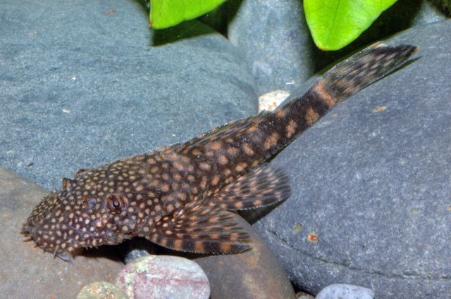
A pair of Jumbie teta, Ancistrus cirrhosus (total length: 10–12 cm) used in this study were purchased from SanHo Aquarium (Wonju, Korea).