New Scleronema
Posted: 02 Jun 2023, 17:08
BOCKMANN, F. A., FERRER, J., RIZZATO, P. P., ESGUÍCERO, A. L., DUBOC, L. F., & INGENITO, L. F. (2023). Anatomy, ecology, and behavior of a new species of Scleronema Eigenmann, 1917 (Siluriformes: Trichomycteridae) from coastal drainages in the southern Brazilian Atlantic Rainforest, with comments on the monophyly and phylogeny of the genus. Zootaxa, 5297(1), 1-47.
https://doi.org/10.11646/zootaxa.5297.1.1
https://www.mapress.com/zt/article/view ... a.5297.1.1
https://doi.org/10.11646/zootaxa.5297.1.1
https://www.mapress.com/zt/article/view ... a.5297.1.1
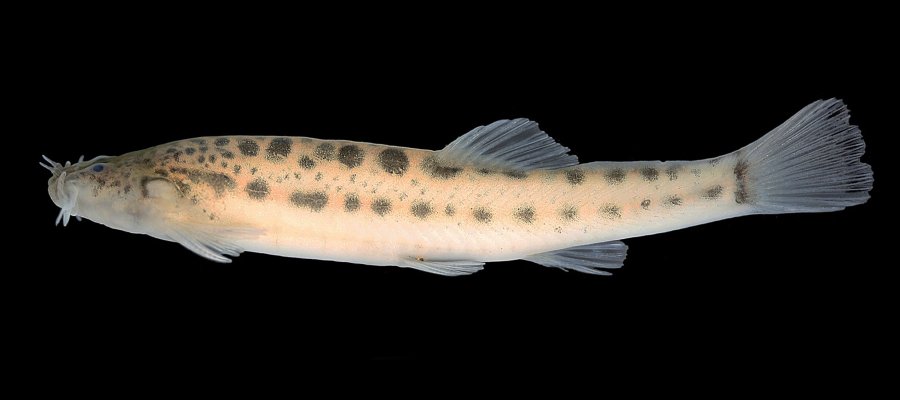
Abstract
A new species of (Trichomycteridae) is described from the lowlands of three coastal river basins in the Atlantic Forest of the Santa Catarina state, Southern Brazil. Aspects of the anatomy, reproduction, diet, feeding behavior and habitat of the new species are described and discussed in comparison with related taxa. The conservation status of the new species, which currently faces several threats due to environmental impacts on its region of occurrence, is established. Based on characteristics observed in the new species, as well as in most of its congeners, the phylogenetic position and monophyly of Scleronema are discussed and traits considered synapomorphic for the subgenera Plesioscleronema and Scleronema are reviewed. The monophyly of the genus Scleronema is supported by a new synapomorphy. In addition, two new synapomorphies, one of which based on behavior, are suggested for the subgenus Scleronema, justifying the inclusion of the new species. Within the subgenus Scleronema, the new species is assigned to the S. minutum group, which currently includes the majority of species of the genus, due to the presence of synapomorphic traits related to the body shape, maxillary barbel, skin flap of the opercle, caudal and pectoral fins, as well as osteological features of the lower jaw, hyoid arch, and postcranial axial skeleton. The species herein described differs from all its congeners by a combination of characters from various morphological complexes, which are described in detail using different methodologies, including radiography, whole-specimen clearing and double-staining procedures, and tridimensional computer nanotomography (3D nano-CT).